|
|
|
Screenshots
The following is a semi-chronological record of significant
milestones in the development of AMIDE.
- 2012.10.17 Demonstrating MPR, MIP, and MINIP rendering modes
for thick slabs (10-mm) from a CT thorax.

- 2005.11.17 Showing off several new featues. The image
display can now be "torn apart" and moved around. The threshold
widget can now use center/width values in addition to min/max
values. Anterior/Posterior/Left/Right/Supine/Inferior
orientations are indicated on the image displays.
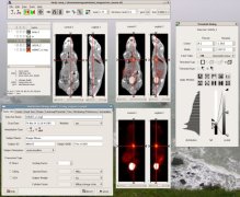
- 2004.01.11 AMIDE (finally) includes a profile tool.

- 2003.05.18 AMIDE now runs under windows too.
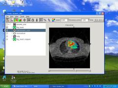
- 2003.03.02 You can now choose which views (transverse,
coronal, and/or sagittal) you want. And a 3-way "linked" mode
can be used in addition to image fusion.

- 2002.10.06 A cropping wizard has been added to allow shrinking of data sets.
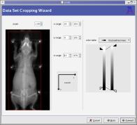
- 2001.12.20 The user can switch between orthogonal and
linear canvas layouts.

- 2001.12.08 Isocontour ROI's have now been added. A 3D
isocontour ROI has been drawn by clicking on the tumor in the
FDG image (hot-metal).
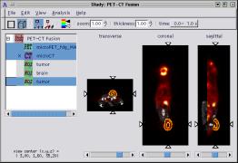
- 2001.11.18 Showing off the new threshold widget, which has
sliders for entering the thresholds as both absolute percentages
(right) and relative percentages (left).
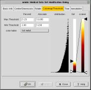
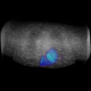
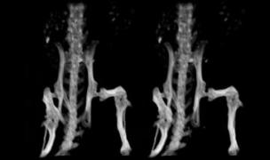
- 2001.10.30 MPEG1 files demonstrating the movie generation
capabilities. The first one is a dynamic FDG scan of the heart
overlayed on the corresponding transmission image. The second
image is a fused microCT and microPET study of a mouse with a
tumor on its right flank. The third one is a stereoscopically
rendered CT image of a mouse, data set courtesy of Andrew Goertzen.

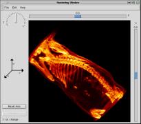
- 2001.10.30 Volume rendering of a mouse CT scan to show off
the new rendering interface. Note that the entire mouse was not
contained in the data set (i.e. it looks like it's headless).
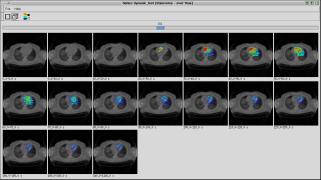
- 2001.10.30 A Time Series of an FDG heart scan (NIH
colorscale) overlayed on a transmission scan (BW colorscale)
showing off the series widget.
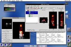
- 2001.10.01 A shot of AMIDE running on Mac OS X. The fink distribution was used to
provide GTK+/GNOME and XonX support. Windowmaker is managing the X windows.
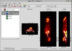
- 2001.08.05 Showing off the new toolbar, which replaces the
widgets which were previously to the right of the canvases. A
merged FDG-PET/CT scan of a mouse is shown. The slice thickness
has been choosen so as to completely enclose the tumor on the
mouse's right flank.
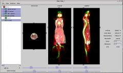
- 2001.01.07 Shows a session containing a superimposed FDG
PET scan (red), CT scan (black/white), and F- PET scan (green)
|

