Advertisement
ArticleImmunology Free access | 10.1172/JCI23913
Virus-induced dysfunction of CD4+CD25+ T cells in patients with HTLV-I–associated neuroimmunological disease
Yoshihisa Yamano,1,2 Norihiro Takenouchi,1 Hong-Chuan Li,3 Utano Tomaru,1,4 Karen Yao,1 Christian W. Grant,1 Dragan A. Maric,5 and Steven Jacobson1
1Viral Immunology Section, Neuroimmunology Branch, National Institute of Neurological Disorders and Stroke, NIH, Bethesda, Maryland, USA. 2Kagoshima City Hospital, Kagoshima, Japan. 3Viral Epidemiology Branch, Division of Cancer Epidemiology and Genetics, National Cancer Institute, Bethesda, Maryland, USA. 4Department of Pathology/Pathophysiology, Hokkaido University Graduate School of Medicine, Sapporo, Japan. 5Laboratory of Neurophysiology, National Institute of Neurological Disorders and Stroke, NIH, Bethesda, Maryland, USA.
Address correspondence to: Steven Jacobson, NIH/NINDS/NIB Building 10, Room 5B-16, Bethesda, Maryland 20892, USA. Phone: (301) 496-0519; Fax: (301) 402-0373; E-mail: [email protected].
Find articles by Yamano, Y. in: JCI | PubMed | Google Scholar
1Viral Immunology Section, Neuroimmunology Branch, National Institute of Neurological Disorders and Stroke, NIH, Bethesda, Maryland, USA. 2Kagoshima City Hospital, Kagoshima, Japan. 3Viral Epidemiology Branch, Division of Cancer Epidemiology and Genetics, National Cancer Institute, Bethesda, Maryland, USA. 4Department of Pathology/Pathophysiology, Hokkaido University Graduate School of Medicine, Sapporo, Japan. 5Laboratory of Neurophysiology, National Institute of Neurological Disorders and Stroke, NIH, Bethesda, Maryland, USA.
Address correspondence to: Steven Jacobson, NIH/NINDS/NIB Building 10, Room 5B-16, Bethesda, Maryland 20892, USA. Phone: (301) 496-0519; Fax: (301) 402-0373; E-mail: [email protected].
Find articles by Takenouchi, N. in: JCI | PubMed | Google Scholar
1Viral Immunology Section, Neuroimmunology Branch, National Institute of Neurological Disorders and Stroke, NIH, Bethesda, Maryland, USA. 2Kagoshima City Hospital, Kagoshima, Japan. 3Viral Epidemiology Branch, Division of Cancer Epidemiology and Genetics, National Cancer Institute, Bethesda, Maryland, USA. 4Department of Pathology/Pathophysiology, Hokkaido University Graduate School of Medicine, Sapporo, Japan. 5Laboratory of Neurophysiology, National Institute of Neurological Disorders and Stroke, NIH, Bethesda, Maryland, USA.
Address correspondence to: Steven Jacobson, NIH/NINDS/NIB Building 10, Room 5B-16, Bethesda, Maryland 20892, USA. Phone: (301) 496-0519; Fax: (301) 402-0373; E-mail: [email protected].
Find articles by Li, H. in: JCI | PubMed | Google Scholar
1Viral Immunology Section, Neuroimmunology Branch, National Institute of Neurological Disorders and Stroke, NIH, Bethesda, Maryland, USA. 2Kagoshima City Hospital, Kagoshima, Japan. 3Viral Epidemiology Branch, Division of Cancer Epidemiology and Genetics, National Cancer Institute, Bethesda, Maryland, USA. 4Department of Pathology/Pathophysiology, Hokkaido University Graduate School of Medicine, Sapporo, Japan. 5Laboratory of Neurophysiology, National Institute of Neurological Disorders and Stroke, NIH, Bethesda, Maryland, USA.
Address correspondence to: Steven Jacobson, NIH/NINDS/NIB Building 10, Room 5B-16, Bethesda, Maryland 20892, USA. Phone: (301) 496-0519; Fax: (301) 402-0373; E-mail: [email protected].
Find articles by Tomaru, U. in: JCI | PubMed | Google Scholar
1Viral Immunology Section, Neuroimmunology Branch, National Institute of Neurological Disorders and Stroke, NIH, Bethesda, Maryland, USA. 2Kagoshima City Hospital, Kagoshima, Japan. 3Viral Epidemiology Branch, Division of Cancer Epidemiology and Genetics, National Cancer Institute, Bethesda, Maryland, USA. 4Department of Pathology/Pathophysiology, Hokkaido University Graduate School of Medicine, Sapporo, Japan. 5Laboratory of Neurophysiology, National Institute of Neurological Disorders and Stroke, NIH, Bethesda, Maryland, USA.
Address correspondence to: Steven Jacobson, NIH/NINDS/NIB Building 10, Room 5B-16, Bethesda, Maryland 20892, USA. Phone: (301) 496-0519; Fax: (301) 402-0373; E-mail: [email protected].
Find articles by Yao, K. in: JCI | PubMed | Google Scholar
1Viral Immunology Section, Neuroimmunology Branch, National Institute of Neurological Disorders and Stroke, NIH, Bethesda, Maryland, USA. 2Kagoshima City Hospital, Kagoshima, Japan. 3Viral Epidemiology Branch, Division of Cancer Epidemiology and Genetics, National Cancer Institute, Bethesda, Maryland, USA. 4Department of Pathology/Pathophysiology, Hokkaido University Graduate School of Medicine, Sapporo, Japan. 5Laboratory of Neurophysiology, National Institute of Neurological Disorders and Stroke, NIH, Bethesda, Maryland, USA.
Address correspondence to: Steven Jacobson, NIH/NINDS/NIB Building 10, Room 5B-16, Bethesda, Maryland 20892, USA. Phone: (301) 496-0519; Fax: (301) 402-0373; E-mail: [email protected].
Find articles by Grant, C. in: JCI | PubMed | Google Scholar
1Viral Immunology Section, Neuroimmunology Branch, National Institute of Neurological Disorders and Stroke, NIH, Bethesda, Maryland, USA. 2Kagoshima City Hospital, Kagoshima, Japan. 3Viral Epidemiology Branch, Division of Cancer Epidemiology and Genetics, National Cancer Institute, Bethesda, Maryland, USA. 4Department of Pathology/Pathophysiology, Hokkaido University Graduate School of Medicine, Sapporo, Japan. 5Laboratory of Neurophysiology, National Institute of Neurological Disorders and Stroke, NIH, Bethesda, Maryland, USA.
Address correspondence to: Steven Jacobson, NIH/NINDS/NIB Building 10, Room 5B-16, Bethesda, Maryland 20892, USA. Phone: (301) 496-0519; Fax: (301) 402-0373; E-mail: [email protected].
Find articles by Maric, D. in: JCI | PubMed | Google Scholar
1Viral Immunology Section, Neuroimmunology Branch, National Institute of Neurological Disorders and Stroke, NIH, Bethesda, Maryland, USA. 2Kagoshima City Hospital, Kagoshima, Japan. 3Viral Epidemiology Branch, Division of Cancer Epidemiology and Genetics, National Cancer Institute, Bethesda, Maryland, USA. 4Department of Pathology/Pathophysiology, Hokkaido University Graduate School of Medicine, Sapporo, Japan. 5Laboratory of Neurophysiology, National Institute of Neurological Disorders and Stroke, NIH, Bethesda, Maryland, USA.
Address correspondence to: Steven Jacobson, NIH/NINDS/NIB Building 10, Room 5B-16, Bethesda, Maryland 20892, USA. Phone: (301) 496-0519; Fax: (301) 402-0373; E-mail: [email protected].
Find articles by Jacobson, S. in: JCI | PubMed | Google Scholar
Published May 2, 2005 - More info
J Clin Invest. 2005;115(5):1361–1368. https://doi.org/10.1172/JCI23913.
© 2005 The American Society for Clinical Investigation
Received: November 16, 2004; Accepted: February 8, 2005
-
Abstract
CD4+CD25+ Tregs are important in the maintenance of immunological self tolerance and in the prevention of autoimmune diseases. As the CD4+CD25+ T cell population in patients with human T cell lymphotropic virus type I–associated (HTLV-I–associated) myelopathy/tropical spastic paraparesis (HAM/TSP) has been shown to be a major reservoir for this virus, it was of interest to determine whether the frequency and function of CD4+CD25+ Tregs in HAM/TSP patients might be affected. In these cells, both mRNA and protein expression of the forkhead transcription factor Foxp3, a specific marker of Tregs, were lower than those in CD4+CD25+ T cells from healthy individuals. The virus-encoded transactivating HTLV-I tax gene was demonstrated to have a direct inhibitory effect on Foxp3 expression and function of CD4+CD25+ T cells. This is the first report to our knowledge demonstrating the role of a specific viral gene product (HTLV-I Tax) on the expression of genes associated with Tregs (in particular, foxp3) resulting in inhibition of Treg function. These results suggest that direct human retroviral infection of CD4+CD25+ T cells may be associated with the pathogenesis of HTLV-I–associated neurologic disease.
-
Introduction
The human T cell lymphotropic virus type I (HTLV-I) is an exogenous human retrovirus that is associated with chronic, persistent infection of human T cells. While the majority of infected individuals remain healthy, lifelong asymptomatic carriers, approximately 2–3% develop an aggressive mature T cell malignancy termed adult T cell leukemia, and another 0.25–3% develop an inflammatory disease of the CNS termed HTLV-I–associated myelopathy/tropical spastic paraparesis (HAM/TSP) (1–3). Furthermore, in some HAM/TSP patients, other autoimmune diseases characterized by multiorgan lymphocytic infiltrates, including uveitis, arthritis, polymyositis, Sjögren syndrome, atopic dermatitis, and alveolitis, have been reported (4, 5). Patients with HAM/TSP have high frequencies of HTLV-I–infected T cells and heightened virus-specific immune responses, including increased proinflammatory cytokine production (6–8). One of the most striking features of the cellular immune response in HAM/TSP patients is the increased numbers of HTLV-I–specific CTLs, which are lower or absent in asymptomatic carriers (9). In some HLA-A*201 HAM/TSP patients, the frequency of Tax11–19–specific CTLs can be as high as 30% of total CD8+ T cells in peripheral blood (10) and even higher in cerebrospinal fluid (6). Neuropathological findings have demonstrated focal infiltrates of T cells and macrophages in the CNS (11). These observations have suggested that inflammatory T cells (particularly virus-specific CD8+ CTLs) may play an immunopathologic role in this disorder.
Recently, a large body of information has demonstrated that CD4+ Tregs constitute an important component of the normal, healthy immune response. These cells are engaged in the maintenance of immunologic self tolerance by actively suppressing the activation and expansion of self-reactive lymphocytes that may cause autoimmune disease (12, 13). The majority of these Tregs constitutively express CD25 (the IL-2 receptor α chain). The normal CD4+CD25+ Treg population constitutes 5–10% of peripheral CD4+ T cells in mice and 1–2% in humans (only the CD4+CD25high T cells exhibit similar regulatory function in humans) (14). Removal or functional alteration of this population from normal rodents leads to the spontaneous development of various autoimmune diseases (12, 13). CD4+CD25+ Tregs have unique immunological characteristics. For example, they do not proliferate in response to antigenic stimulation in vitro and can potently suppress the proliferation of other CD4+ or CD8+ T cells induced either by polyclonal or antigen-specific stimuli (12, 13). Costimulation with anti-CD28 or provision of exogenous IL-2 inhibits the suppressive ability of these CD4+CD25+ Tregs (15, 16). They constitutively express gene products of glucocorticoid-induced TNF receptor family–related (GITR) receptors and cytotoxic T lymphocyte–associated antigen 4 (CTLA-4) (17–21). Furthermore, it has been reported that forkhead transcription factor (foxp3) gene is specifically expressed in Tregs and is required for their development and function (22–24). Interestingly, mice of the foxp3 mutant strain, or scurfy mice, succumb to a CD4+ T cell–mediated, lymphoproliferative, and autoimmune disease characterized by multiorgan lymphocytic infiltrates and overproduction of proinflammatory cytokines (25–27). Furthermore, similar immunological abnormalities are observed in CTLA-4–deficient mice (28, 29). HAM/TSP patients share many immunological characteristics with the scurfy foxp3 mutants and CTLA-4–deficient mice, including the in vitro spontaneous lymphoproliferation of predominantly CD4+ T cells and clinical manifestations associated with autoimmune disease characterized by multiorgan lymphocytic infiltrates and overproduction of proinflammatory cytokines. It was therefore of interest to determine the frequency and function of CD4+ Tregs in patients with HAM/TSP.
We have recently demonstrated that in HAM/TSP patients, the CD4+CD25+ T cell population is the main reservoir for HTLV-I: more than 90% of these cells contain HTLV-I proviral DNA, and they express HTLV-I tax mRNA at significantly higher levels than in CD4+CD25– cells (30). Moreover, these HTLV-I–infected CD4+CD25+ T cells were not functionally suppressive but rather were shown to be stimulatory for the HTLV-I Tax–specific proliferation of CD8+ T cells (30). Therefore, we have hypothesized that HTLV-I infection of CD4+CD25+ T cells may alter the regulatory function of this population of CD4+ cells or that the proportion of Tregs may be decreased in HAM/TSP patients. To answer these questions, we developed a quantitative TaqMan PCR assay for the detection of human foxp3 mRNA and a FACS assay for the detection of Foxp3 protein. We have shown that foxp3 mRNA expression in CD4+CD25+ T cells of HAM/TSP patients is lower than that of HDs. In addition, CD4+CD25+ T cells of HAM/TSP patients have lower levels of expression of Foxp3 protein as well as other Treg markers such as CTLA-4 and GITR but were overproducing proinflammatory cytokines such as IL-2 that are known to inhibit CD4+CD25+ regulatory activity. Importantly, we have also demonstrated defects in the regulatory function of HTLV-I tax gene–transfected CD4+CD25+ T cells. In an attempt to define which HTLV-I virus gene(s) may be associated with the dysregulation of Foxp3, we have transfected the HTLV-I–transactivating tax gene into CD4+CD25+ T cells from HDs and have demonstrated a Tax-specific inhibition of foxp3 expression that can suppress CD4+CD25+ Treg function. Collectively, these results demonstrate that a consequence of HTLV-I infection of CD4+CD25+ T cells in HAM/TSP patients (30) is the suppression in both the frequency and function of CD4+ Tregs, which may be associated with a break in immunological self tolerance resulting in the HTLV-I–associated disorders with multiorgan lymphocytic infiltrates.
-
Results
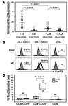
Decreased foxp3 expression in CD4+CD25+ T cells from HAM/TSP patients. To assess whether CD4+CD25+ cells in HAM/TSP patients have altered expression of Foxp3, we isolated CD4+CD25+ and CD4+CD25- T cells from PBMCs of HAM/TSP patients, HTLV-I–infected asymptomatic carriers (ACs), and uninfected healthy donors (HDs) and quantified the expression levels of foxp3 by real-time RT-PCR. The percentages (mean ± SD) of CD4+CD25high T cells in PBMCs of HAM/TSP patients, ACs, and HDs were 19.52% ± 9.00%, 5.30% ± 1.62%, and 2.19% ± 1.07%, respectively (Supplemental Figure 1; supplemental material available online with this article; doi:10.1172/JCI200523913DS1). As expected, foxp3 mRNA expression levels were significantly higher (P = 0.0015) in CD4+CD25+ cells compared with CD4+CD25– cells from 13 HDs (Figure 1A). Similarly, foxp3 expression levels were also higher in CD4+CD25+ cells compared with CD4+CD25– T cells from 13 HAM/TSP patients (P = 0.0024). However, the expression of foxp3 in the HAM/TSP CD4+CD25+ population (6.81 ± 4.77; see Methods) was significantly lower (approximately 2.5-fold; P = 0.0011) than that observed in HD CD4+CD25+ cells (16.01 ± 10.76; see Methods) (Figure 1A). foxp3 expression levels in CD4+CD25+ cells from 2 ACs were comparable to levels observed in cells from HDs (Table 1). No difference in the expression levels of foxp3 mRNA was observed among HAM/TSP, AC, and HD CD4+CD25– cells. These results are in agreement with previous studies of both mouse and human (22, 31) Tregs demonstrating that the transcription factor Foxp3 is preferentially expressed in CD4+CD25+ T cells. However, the foxp3 expression was reduced in CD4+CD25+ T cells from patients with HAM/TSP.
 Figure 1
Decreased Foxp3 expression in CD4+CD25+ T cells from HAM/TSP patients. (A) Quantitative expression of foxp3 mRNA was determined by real-time RT-PCR. The level of foxp3 mRNA expression was calculated as the relative quantity of foxp3 mRNA expression divided by the relative quantity of endogenous control HPRT mRNA expression, as described in Methods. The data represent isolated cell subsets (CD4+CD25+ or CD4+CD25–) from 13 uninfected HDs and 13 HAM/TSP patients (HAM). Foxp3 mRNA expression was significantly reduced in the CD4+CD25+ T cell subset from HDs compared with that from HAM/TSP patients. (B) A representative histogram of intracellular expression of Foxp3 protein showing results from flow cytometric analysis of PBMC samples from HAM/TSP patients and HDs. Foxp3 protein expression was detected in the CD4+CD25+ T cell subset from HDs but not in CD4+CD25– or in total CD4– T cell subsets. In contrast, the number of Foxp3-positive cells in CD4+CD25+ T cells from HAM/TSP patients was clearly reduced. (C) Data represent averaged percentage of Foxp3-positive cells in each T cell subset. The percentage (mean ± SD) of Foxp3-positive cells in CD4+CD25+ T cells of 8 HAM/TSP patients (3.09% ± 1.04%) was significantly lower than that of 8 HDs (25.9% ± 8.23%; P = 0.0014). No difference in the protein expression levels of Foxp3 was observed in CD4+CD25– or CD4– cells between HAM/TSP patients and HDs.
 Table 1
foxp3 mRNA expression in CD4+CD25+ T cells and CD4+CD25– T cells from HAM/TSP patients, ACs, and HDs
Loss of foxp3 protein expression on CD4+CD25+ T cells from HAM/TSP patients. As we had shown that the level of foxp3 mRNA was significantly decreased in CD4+CD25+ T cells from HAM/TSP patients compared with HDs, we wished to determine whether comparable reductions in Foxp3 protein expression could also be demonstrated. Therefore, we investigated the intracellular expression of Foxp3 protein in PBMCs from HAM/TSP patients and HDs using flow cytometry with a commercially available anti-human Foxp3 antibody. Analysis of Foxp3 protein expression in subpopulations of lymphocytes from 8 HDs revealed significant staining, as expected, in the CD4+CD25+ T cell subset but not the CD4+CD25– or CD4– T cell subsets (Figure 1, B and C). A representative histogram is shown in Figure 1B. The percentage (mean ± SD) of Foxp3-positive cells in CD4+CD25+ T cells from 8 HDs was 25.9% ± 8.23% (Figure 1C). This is consistent with the hypothesis that only a subset of the CD4+CD25+ T cell population may be CD4+ Tregs (12, 13). In contrast, the percentage (mean ± SD) of Foxp3-positive cells in CD4+CD25+ T cells from 8 HAM/TSP patients was significantly reduced to 3.09% ± 1.04% (P = 0.0014) (Figure 1C). A representative histogram is shown in Figure 1B. No difference in the protein expression levels of Foxp3 was observed in CD4+CD25– or CD4– cells between HAM/TSP patients and HDs (Figure 1B). These results support the finding that foxp3 mRNA is reduced in CD4+CD25+ cells from HAM/TSP patients compared with HDs (Figure 1A) and continue to suggest that dysregulation of Tregs may be contribute to the pathogenesis of this disorder.
Reduced expression of regulatory cell surface marker and increased proinflammatory cytokine production in CD4+CD25+ T cells from HAM/TSP patients. Tregs have been characterized by their constitutive expression not only of Foxp3 but also of cell surface proteins such as CD25, CD38, CD62L, CD69, CTLA-4, and GITR (17–21, 23, 32, 33). To determine the levels of these cell surface molecules, we investigated their expression in CD4+CD25+ T cells from both HAM/TSP patients and HDs. As shown in Table 2, CD4+CD25+ T cells from HAM/TSP patients showed lower expression of CD38 (P = 0.0003), CD62L (P = 0.0374), CD69 (P = 0.0101), CTLA-4 (P = 0.0104), and GITR (P = 0.0010) molecules than those from HDs, while the expression of HLA-DR was not significantly different. We confirmed a decrease in CD45RA expression (P = 0.0112) and an increase in CD45RO expression (P < 0.0001) in CD4+CD25+ T cells from HAM/TSP patients (Table 2), as had been previously reported (34, 35). We also investigated intracellular cytokine expression in CD4+CD25+ T cells. The expression of proinflammatory cytokine such as IL-2 (P = 0.0011) and IFN-γ (P = 0.0034) was significantly increased in HAM/TSP patients compared with HDs, whereas there were no significant differences in expression of Th2 cytokines such as IL-4 and IL-10 (Table 2). Collectively, these results demonstrate a reduction in cell surface molecules, particularly GITR and CTLA-4, which have been associated with CD4+ Tregs, on HAM/TSP CD4+CD25+ cells (17–21). These findings are consistent with our previous observations on reduced Foxp3 expression (Figure 1).
 Table 2
Cell surface marker expression and proinflammatory cytokine production in CD4+CD25+ T cells from HAM/TSP patients and HDs
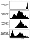
Lack of regulatory function in CD4+CD25+ T cells from HAM/TSP patients. While we have shown a decrease in foxp3 mRNA and protein expression in HAM/TSP CD4+CD25+ cells as well as other cell surface markers that characterize CD4+ Tregs, it remains to be determined whether this corresponds to a reduction in Treg function. To determine the effect of HAM/TSP CD4+CD25+ cells on T cell regulatory function, we performed functional CFSE proliferation assays. As shown Figure 2, HD CD4+CD25– T cells specifically proliferated upon stimulation with anti-CD3 antibody. As expected, addition of irradiated, sorted allogeneic HD CD4+CD25+ (which did not proliferate; data not shown) to these HD CD4+CD25–-responding cells resulted in an inhibition of proliferation consistent with a Treg function of HD CD4+CD25+ cells (14, 36, 37). In contrast, coculturing irradiated HAM/TSP CD4+CD25+ cells with HD CD4+CD25– cells did not suppress the proliferative capacity of these anti-CD3–stimulated, responding CD4+CD25– cells (Figure 2). These results suggest that Treg function in CD4+CD25+ cells from HAM/TSP patients is dysregulated.
 Figure 2
Lack of regulatory function in CD4+CD25+ T cells from HAM/TSP patients. A total of 1 × 10⁵ CD4+CD25– T cells/well from HDs were labeled with CFSE. They were cultured for 6 days in the culture medium in the absence or presence of 2.5 μg/ml anti-CD3 antibody (top 2 panels). They were also cultured for 6 days in 2.5 μg/ml anti-CD3 antibody added to culture medium with 1 × 10⁵ irradiated allogeneic CD4+CD25+ T cells from HDs or with 1 × 10⁵ irradiated CD4+CD25+ T cells from HAM/TSP patients (bottom 2 panels). The data indicate that regulatory function in CD4+CD25+ T cells from HAM/TSP patients is reduced in comparison with that in CD4+CD25+ T cells from HDs. Failure of CD4+CD25+ T cells to suppress lymphoproliferation of activated HD cells was observed in separate experiments with cells from 4 HAM/TSP patients, while suppression of activated HD cell proliferation by allogeneic HD CD4+CD25+ T cells from 2 HDs was demonstrated.
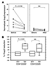
HTLV-I Tax suppresses foxp3 expression. Since Foxp3 message and protein expression were significantly reduced in HAM/TSP CD4+CD25+ cells relative to those from HDs, we hypothesized that the virus-encoded transactivating tax gene (38, 39) might be associated with this reduction. To investigate this possibility, we transfected an HTLV-I tax DNA vector known to express high levels of HTLV-I Tax protein (40) into purified CD4+CD25+ T cells and CD4+CD25– T cells from 7 HDs using a highly efficient electroporation transfection system (greater than 70% of transfected cells expressed the transgene). We measured foxp3 mRNA expression using real-time RT-PCR before and after transfection. As shown in Figure 3, in all donors, the foxp3 mRNA expression level in CD4+CD25+ T cells was significantly decreased by transfection with HTLV-I tax DNA (P = 0.018). By contrast, there was no significant difference in the level of foxp3 message in CD4+CD25– T cells before and after HTLV-I tax DNA transfection (Figure 3, A and B). When CD4+CD25+ T cells from HDs were transfected with another HTLV-I gene expression vector, HTLV-I env, no change in foxp3 mRNA expression level was observed (Figure 3B). These results support the hypothesis that the transactivating HTLV-I tax gene is associated with the reduction in foxp3 message and protein expression observed in HAM/TSP CD4+CD25+ T cells.
 Figure 3
HTLV-I Tax suppresses Foxp3 expression. Purified CD4+CD25+ T cells and CD4+CD25– T cells from HDs were transfected with the HTLV-I tax gene (n = 7) or HTLV-I env gene (n = 4). The foxp3 mRNA expression in these T cell populations before and after transfection was measured by real-time RT-PCR. (A) The foxp3 mRNA expression level in CD4+CD25+ T cells was significantly decreased by transfection with HTLV-I tax gene (P = 0.018). By contrast, there was no significant decrease in foxp3 mRNA expression in CD4+CD25– T cells. (B) foxp3 mRNA expression was significantly decreased in HTLV-I tax–transfected CD4+CD25+ T cells compared with HTLV-I env–transfected CD4+CD25+ T cells (P = 0.020). There was no significant difference between the foxp3 mRNA expression in HTLV-I tax transfected CD4+CD25– T cells and that in HTLV-I env–transfected CD4+CD25– T cells. env, HTLV-I env gene; tax, HTLV-I tax gene.
Loss of regulatory function in HTLV-I tax–transfected HD CD4+CD25+ T cells. As we had demonstrated that HTLV-I tax significantly reduced foxp3 messenger RNA levels in HTLV-I tax–transfected HD CD4+CD25+ cells, it was of interest to determine whether this also corresponded to a reduction in T cell regulatory function in this population of cells. As shown in Figure 4 (a representative experiment using cells from 3 different HDs), HD CD4+CD25– T cells alone proliferated upon stimulation with anti-CD3 antibody, while the capacity of HD CD4+CD25+ regulatory cells to proliferate upon this stimulus was significantly diminished. As expected, addition of HD CD4+CD25+ to autologous HD CD4+CD25–-responding cells demonstrated an inhibition of proliferation. In contrast, coculturing of HTLV-I Tax–transfected HD CD4+CD25+ cells (which induced a reduction in foxp3 message; Figure 3) with HD CD4+CD25– failed to suppress the proliferation of these anti-CD3–stimulated, responding CD4+CD25– cells (Figure 4). These results support the hypothesis that the reduction of levels in Foxp3 in HAM/TSP CD4+CD25+ cells is mediated through infection with HTLV-I and may result in dysregulation in Treg function of HTLV-I–infected CD4+CD25+ Tregs.
![Loss of regulatory function in HTLV-I tax–transfected HD CD4+CD25+ T cells. CD4+CD25+ or CD4+CD25– T cells from uninfected HDs were stimulated with 2.5 μg/ml anti-CD3 antibody and irradiated PBMCs and cultured for 4 days (HD CD25+ and HD CD25–). Furthermore, to compare the suppressive activity of HD CD4+CD25+ T cells before and after HTLV-I tax gene transfection, CD4+CD25– T cells from HDs were stimulated with 2.5 μg/ml anti-CD3 antibody and irradiated PBMCs and cultured for 4 days in the presence of equal numbers of HD CD4+CD25+ T cells or HTLV-I tax–transfected HD CD4+CD25+ T cells (Tax+ HD CD25+). After culture, [3H]thymidine was added for additional 16 hours. The suppressive activity of CD4+CD25+ T cells from HDs was inhibited by transfection with the HTLV-I tax gene. Data represent the mean of experiments with cells from 3 HDs. Loss of regulatory function in HTLV-I tax–transfected HD CD4+CD25+ T cells.](//dm5migu4zj3pb.cloudfront.net/manuscripts/23000/23913/small/JCI0523913.f4.gif) Figure 4
Figure 4Loss of regulatory function in HTLV-I tax–transfected HD CD4+CD25+ T cells. CD4+CD25+ or CD4+CD25– T cells from uninfected HDs were stimulated with 2.5 μg/ml anti-CD3 antibody and irradiated PBMCs and cultured for 4 days (HD CD25+ and HD CD25–). Furthermore, to compare the suppressive activity of HD CD4+CD25+ T cells before and after HTLV-I tax gene transfection, CD4+CD25– T cells from HDs were stimulated with 2.5 μg/ml anti-CD3 antibody and irradiated PBMCs and cultured for 4 days in the presence of equal numbers of HD CD4+CD25+ T cells or HTLV-I tax–transfected HD CD4+CD25+ T cells (Tax+ HD CD25+). After culture, [3H]thymidine was added for additional 16 hours. The suppressive activity of CD4+CD25+ T cells from HDs was inhibited by transfection with the HTLV-I tax gene. Data represent the mean of experiments with cells from 3 HDs.
-
Discussion
Naturally arising CD4+CD25+ Tregs are engaged in dominant control of self-reactive T cells, contributing to the maintenance of immunological self tolerance. It has been known that foxp3 is specifically expressed in CD4+CD25+ Tregs and is a key gene for the development and function of Tregs (22–24). Therefore, to test the hypothesis that HTLV-I–infected CD4+CD25+ T cells may lack regulatory potential in HAM/TSP patients, we measured foxp3 gene expression quantitatively and demonstrated that Foxp3 expression in CD4+CD25+ T cells of HAM/TSP patients was lower than that in cells of HDs (Figure 1). This result suggested 3 possibilities: HTLV-I has a direct inhibitory effect on Foxp3 expression; the frequency of Tregs is decreased in the CD4+CD25+ T cell population of HAM/TSP patients; or HAM/TSP patients have genetically determined low expression of foxp3 gene. Although these possibilities are not mutually exclusive, to address whether HTLV-I has direct inhibitory effect on the Foxp3 expression, we tested the effect of HTLV-I tax gene transfection on foxp3 expression in CD4+CD25+ T cells from HDs. As shown in Figures 3 and 4, it was demonstrated that HTLV-I Tax had a direct inhibitory effect on Foxp3 expression and inhibited the regulatory function of CD4+CD25+ T cells from HDs. These results suggest that HTLV-I has the potential to induce the diminution of CD4+CD25+ Treg function through the suppression of Foxp3 expression. Moreover, this is the first report to our knowledge demonstrating the role of a specific viral gene product (HTLV-I Tax) on the expression of Foxp3 that results in inhibition of Treg function. Potentially other viruses tropic for CD4+ cells may have similar effects on this important function of Tregs, as has been recently reported for HIV (41, 42).
The analysis of cell surface markers and cytokine production of CD4+CD25+ T cells from HAM/TSP further supports the observations of reduced Foxp3 levels. CD4+CD25+ T cells from HAM/TSP patients expressed lower levels of CTLA-4 and GITR molecules. CTLA-4 and GITR have also been reported to be constitutively expressed on Tregs and play a key role in normal CD4+CD25+ Treg function (17–21). Therefore, reduced expression of CTLA-4 and GITR on CD4+CD25+ T cells of HAM/TSP patients may suggest decreased frequency of Tregs in HAM/TSP patients. However, as the CTLA-4 expression on CD4+CD25+ T cells is not decreased in scurfy foxp3 mutant mice (23), this low expression of CTLA-4 on CD4+CD25+ T cells of HAM/TSP patients is not caused by low Foxp3 expression. HTLV-I may have direct suppressive effect on CTLA-4 expression. Furthermore, CD4+CD25+ T cells from HAM/TSP patients overproduced proinflammatory cytokines such as IL-2 and IFN-γ (Table 2) and may contribute to the spontaneous lymphoproliferation that has been observed in such patients (43, 44). It has been reported that normal CD4+CD25+ Tregs do not produce IL-2 by themselves and lose regulatory function in the presence of exogenous IL-2 (15, 16). Therefore, increased production of IL-2 may further support the hypothesis that CD4+CD25+ T cells from HAM/TSP patients have a defect in Treg function.
Activated T cells are increased in HAM/TSP patients (Table 2), and this raises the possibility that there may be a dilution of Tregs rather than a functional decrease in this population. To minimize this concern, we selected the CD25+ population from HAM/TSP patients based on gates set on CD25high in HDs during FACS sorting. A number of studies have shown that predominantly CD25high T cells possess regulatory functions, while CD25low represent activated T cells (14, 37, 45). Importantly, we have direct evidence that the introduction of HTLV-I tax downregulated foxp3 expression in HD CD4+CD25+ T cells, while HTLV-I env did not (Figure 3B). This downregulation of foxp3 was associated with a decrease in Treg function (Figure 4).
To demonstrate functional dysregulation, we also compared the ability of CD4+CD25+ Tregs isolated from HDs and from HAM/TSP patients to suppress plate-bound CD3-activated CD4+CD25– T cells from HDs. As shown in Figures 2 and 4, proliferation of plate-bound CD3-activated CD4+CD25– cells was diminished by 30% (Figure 4) with HD CD4+CD25+ T cells, while HAM/TSP CD4+CD25+ T cells (Figure 2) or HTLV-I tax–transfected T cells (Figure 4) did not suppress T cell proliferation. Collectively, these data suggest defects in the function of HAM/TSP CD4+CD25+ Tregs. The suppression of activated CD4+CD25– T cells by Tregs we observed is consistent with previous reports (12, 14, 16, 46), although Baecher-Allan et al. have demonstrated inhibition of CD4+CD25+ Treg function when responding CD4+CD25– cells were stimulated with high concentrations of plate-bound CD3 (46). Difference in these 2 studies could be explained by the different ratios of responding suppressor T cells used. In the present study, we demonstrated the suppressive function using a 1:1 ratio of CD4+CD25+ Tregs to responder cells.
It has been reported that naturally present Tregs may act to hamper effective immune responses to invading pathogenic microbes (33, 47, 48). For example, in mice infected with Friend retrovirus, it was demonstrated that CD4+ Tregs were increased in number and showed immunosuppressive activity. These CD4+ Tregs had increased expression of CD38+ and CD69+ (33). In contrast, the expression of CD38+ and CD69+ on CD4+CD25+ T cells was decreased in HAM/TSP patients and did not show immunosuppressive activity. These results suggest that CD38+ and CD69+ are also important cell surface markers that may distinguish human Tregs from effector T cells, as reported previously in studies on rodents (32, 33).
It has been reported that microbial infection can dysregulate Tregs to suppress pathologic antimicrobial immune responses that cause tissue damage (i.e., immunopathologic response) (49, 50). For example, in SCID mice chronically infected with Pneumocystis carinii, transfer of T cells depleted of CD4+CD25+ Tregs elicited severe pneumonitis, whereas transfer of T cells not depleted of Tregs did not (49). Thus, in controlling microbial immunity, the frequency of CD4+CD25+ Tregs may play an important role. However, it is not known how these T cells contribute to the regulation of antimicrobial immune responses. The increased expression of CD28 molecules and decreased expression of CTLA-4 on CD4+CD25+ T cells in HAM/TSP patients (shown in this study) may therefore serve to regulate this population of cells (51). CD28 and CTLA-4 share the same ligands (CD80 and CD86) on APCs, and CD28 has much lower affinity for CD80 and CD86 than CTLA-4 (52). CTLA-4 has been reported to be required for the suppressive function of Tregs. In contrast, stimulation through CD28, with concurrent TCR stimulation, abrogates suppressive function (15, 16). CD4+CD25+ T cells have been reported to be a major reservoir of HTLV-I and to present HLA-virus peptide complexes (30). This increased expression of HTLV-I peptide/HLA complexes on CD4+CD25+ cells may increase activation of these cells by signaling through CD28, resulting in the loss of T cell regulatory/suppressive activity. Further comparative analysis of the expression of these molecules on CD4+CD25+ T cells between healthy individuals infected with HTLV-I and patients with HAM/TSP will be necessary to confirm these hypothesis.
In summary, it was demonstrated that in CD4+CD25+ T cells from HAM/TSP patients that were preferentially infected with HTLV-I, Foxp3 expression was lower than that in cells from HDs. HTLV-I Tax had a direct inhibitory effect on Foxp3 expression and inhibited the regulatory function of CD4+CD25+ T cells from HDs. Furthermore, compared to CD4+CD25+ T cells from HD, CD4+CD25+ T cells from HAM/TSP patients showed lower expression of constitutive molecules of Tregs such as CD38, CD62L, CD69, CTLA-4, and GITR and overproduced proinflammatory cytokines such as IL-2 and IFN-γ. In addition, loss of function of CD4+CD25+ T cells has also been reported in other autoimmune disorders such as type 1 diabetes, rheumatoid arthritis, and multiple sclerosis, a neurodegenerative disorder of unknown etiology (37, 45, 53). The finding that autoreactive T cells in patients with autoimmune diseases are more easily activated (54, 55) than those in healthy individuals suggest that CD4+CD25+ Tregs may play a role in controlling the development of autoimmunity. A dysfunction in Tregs in HAM/TSP is consistent with the hypothesis that an autoimmune component may also contribute to the pathogenesis of HAM/TSP (reviewed in ref. 56). Although it has been well demonstrated that the removal or functional alteration of CD4+CD25+ Tregs from normal rodents leads to the spontaneous development of autoimmune diseases, how these cells lose their suppressive function in human disease is unknown. This study suggests the hypothesis that the direct human retrovirus infection of CD4+CD25+ T cells may contribute to a dysregulation of CD4+CD25+ Tregs in a human retro-virus-associated neurologic disease.
-
Methods
Subjects and cell preparation. The PBMCs were prepared by centrifugation over Ficoll-Hypaque gradients (BioWhittaker) from 13 HAM/TSP patients, 13 HTLV-I–seronegative HDs, and 2 ACs, and the cells were viably cryopreserved in liquid nitrogen until tested. HAM/TSP was diagnosed according to WHO guidelines (57). HTLV-I seropositivity was determined by ELISA (Abbott Laboratories), with confirmation by Western blot analysis (Genelabs Technologies Inc.). Blood samples were obtained after informed consent as part of a clinical protocol reviewed and approved by the NIH institutional review panel. CD4+ T cells were negatively selected from the PBMCs with magnetic beads (MACS CD4+ T cell isolation kit; Miltenyi Biotec) according to the manufacturer’s instructions. These selected CD4+ T cells were stained with anti-CD25 FITC (Caltag Laboratories) and sorted into CD4+CD25+ (sorted CD25+ cells were gated on high levels of expression of CD25 in HDs during FACS sorting; Supplemental Figure 1) and CD4+CD25– T cells using FACSVantage (BD).
Foxp3 expression analysis by real-time RT-PCR. Total RNA was extracted using RNeasy Mini Kit (QIAGEN) according to the manufacturer’s instructions, and cDNA was synthesized from extracted RNA using TaqMan Gold RT-PCR Kit using Random Hexamer primer (Applied Biosystems). foxp3 mRNA expression was quantified by real-time PCR using ABI PRISM 7700 Sequence Detector (Applied Biosystems). Real-time RT-PCR was performed using the protocol described in our previous report (10), with some modification. Sample cDNA from 100 ng RNA was applied per well and analyzed. Samples were run in duplicate, and the mean values were used for calculation. The primer set for foxp3 was 5′-GGCCCTTCTCCAGGACAGA-3′ and 5′-GCTGATCATGGCTGGGTTGT-3′. The probe for foxp3 was 5′-FAM-ACTTCATGCATCAGCTCTCCACTGTGGAT-TAMRA-3′. Amplification was carried out at 50°C for 2 minutes, 95°C for 10 minutes, and 45 cycles at 95°C for 15 seconds and 60°C for 1 minute in a total volume of 50 μl. We used the human housekeeping gene hypoxanthine ribosyl transferase (HPRT) primers and probe set (Applied Biosystems) to calculate for normalized values of foxp3 mRNA expression. The normalized values in each sample were calculated as the relative quantity of foxp3 mRNA expression divided by the relative quantity of HPRT mRNA expression. The values were calculated by the following formula: normalized foxp3 expression = 2Ct value of HPRT — Ct value of foxp3.
Flow cytometric analysis. PBMCs were immunostained with various combinations of the following fluorescence-conjugated antibodies: CD25 (CALTAG Laboratories), CD4, CD45RA, CD45RO, CD27, CD28, CD38, CD62L, HLA-DR, CD69, CTLA-4 (BD Biosciences — Pharmingen), and GITR (R&D Systems). These cells were also intracellularly stained with the following antibodies: IL-2, IFN-γ, IL-4, IL-10 (BD Biosciences — Pharmingen), and Foxp3 (Abcam Inc). Flow cytometric analysis was performed on a FACSCalibur cytometer (BD Biosciences). Data processing was accomplished with CELLQuest software (BD).
Transfection. The sorted cells from HDs were harvested in a seeding condition of 1 × 10⁶ cell/ml and were incubated for 2 hours at 37°C in RPMI 1640 supplemented with 10% FCS, 100 μg/ml streptomycin, 100 U/ml penicillin, and 2 mM glutamine (culture medium). The cells washed once in PBS and resuspended in the specified electroporation buffer (Nucleofector solution; Amaxa) to a final concentration of 1 × 10⁶ cells/100 μl. Then 2 μg of HTLV-I env plasmid DNA or HTLV-I tax plasmid DNA (kindly provided by D. Derse, National Cancer Institute–Frederick, Frederick, Maryland, USA) were added to the cell suspension and they were transfected using T cell Nucleofector kit (Amaxa) according to the manufacturer’s instructions. After electroporation, the cells were immediately suspended in 2 ml of culture medium and cultured overnight at 37°C in a 5% CO2 incubator.
Proliferation assay by CFSE. A total of 1 × 10⁵ CD4+CD25– T cells/well from HD were labeled with CFSE using Vybrant CFDA SE Cell Tracer Kit (Invitrogen Corp.) according to the manufacturer’s instructions. Cells were incubated for 6 days in the culture medium with or without 2.5 μg/ml anti-CD3 antibody in round-bottomed 96-well plates. In some cultures, 1 × 10⁵ irradiated allogeneic CD4+CD25+ T cells from HDs or 1 × 10⁵ irradiated allogenic CD4+CD25+ T cells from HAM/TSP patients were added. Cells were subjected to flow cytometric analysis.
Proliferation assay by liquid scintillation counter. For the proliferation assay of T cells from HDs, 1 × 10⁴ CD4+CD25+ or CD4+CD25– T cells/well from HDs were cultured in 200 μl culture medium (RPMI 1640 supplemented with L-glutamine, penicillin, streptomycin, and 5% human AB serum) in round-bottomed 96-well plates. These cell populations were stimulated with 2.5 μg/ml anti-CD3 antibody (OKT-3; BD) in the presence of 5 × 10⁴ irradiated PBMCs. After 4 days culture, 1 μCi tritium thymidine ([3H]TdR)/well was added for additional 16 hours. A liquid scintillation counter was used to measure proliferation. Furthermore, to compare the suppressive effect on the cell proliferation between CD4+CD25+ T cells and HTLV-I tax gene transfected CD4+CD25+ T cells, 1 × 10³ CD4+CD25– T cells/well from HD were stimulated with 2.5 μg/ml anti-CD3 antibody (OKT-3) in the presence of 5 × 10⁴ irradiated PBMCs, then cocultured with 1 × 10⁴ CD4+CD25+ T cells/well or with 1 × 10⁴ HTLV-I tax–transfected CD4+CD25+ T cells/well. After 4 days culture, 1 μCi [3H]TdR/well was added for additional 16 hours. A liquid scintillation counter was used to measure proliferation.
Statistical analysis. Student’s t tests were used for the significance of data comparison.
-
Footnotes
See the related Commentary beginning on page 1144.
Nonstandard abbreviations used: AC, HTLV-I–infected asymptomatic carrier; GITR, glucocorticoid-induced TNF receptor family–related; HAM/TSP, HTLV-I–associated myelopathy/tropical spastic paraparesis; HD, healthy donor; HTLV-I, human T cell lymphotropic virus type I.
Conflict of interest: The authors have declared that no conflict of interest exists.
-
References
- Osame, M, et al. HTLV-I associated myelopathy, a new clinical entity [letter]. Lancet. 1986. 1:1031-1032. View this article via: PubMed Google Scholar
- Gessain, A, et al. Antibodies to human T-lymphotropic virus type-I in patients with tropical spastic paraparesis. Lancet. 1985. 2:407-410.
- Kaplan, JE, et al. The risk of development of HTLV-I-associated myelopathy/tropical spastic paraparesis among persons infected with HTLV-I. J. Acquir. Immune. Defic. Syndr. 1990. 3:1096-1101. View this article via: PubMed Google Scholar
- Nakagawa, M, et al. HTLV-I-associated myelopathy: analysis of 213 patients based on clinical features and laboratory findings. J. Neurovirol. 1995. 1:50-61.
- Uchiyama, T. Human T cell leukemia virus type I (HTLV-I) and human diseases. Annu. Rev. Immunol. 1997. 15:15-37.
- Nagai, M, Yamano, Y, Brennan, MB, Mora, CA, Jacobson, S. Increased HTLV-I proviral load and preferential expansion of HTLV-I Tax-specific CD8+ T cells in cerebrospinal fluid from patients with HAM/TSP. Ann. Neurol. 2001. 50:807-812.
- Jacobson, S. Immunopathogenesis of human T cell lymphotropic virus type I-associated neurologic disease. J. Infect. Dis. 2002. 186(Suppl. 2):187-192. View this article via: CrossRef Google Scholar
- Osame, M. Pathological mechanisms of human T-cell lymphotropic virus type I-associated myelopathy (HAM/TSP). J. Neurovirol. 2002. 8:359-364.
- Jacobson, S, Shida, H, McFarlin, DE, Fauci, AS, Koenig, S. Circulating CD8+ cytotoxic T lymphocytes specific for HTLV-I pX in patients with HTLV-I associated neurological disease. Nature. 1990. 348:245-248.
- Yamano, Y, et al. Correlation of human T-cell lymphotropic virus type 1 (HTLV-1) mRNA with proviral DNA load, virus-specific CD8(+) T cells, and disease severity in HTLV-1-associated myelopathy (HAM/TSP). Blood. 2002. 99:88-94.
- Izumo, S., et al. 1989. The neuropathology of HTLV-I associated myelopathy in Japan: report of an autopsy case and review of the literature. In HTLV-I and the nervous system. G.C. Roman, J.C. Vernant, and M. Osame, editors. Alan R. Liss. New York, New York, USA. 261–267.
- Sakaguchi, S, et al. Immunologic tolerance maintained by CD25+ CD4+ regulatory T cells: their common role in controlling autoimmunity, tumor immunity, and transplantation tolerance. Immunol. Rev. 2001. 182:18-32.
- Shevach, EM. CD4+ CD25+ suppressor T cells: more questions than answers. Nat. Rev. Immunol. 2002. 2:389-400. View this article via: PubMed Google Scholar
- Baecher-Allan, C, Brown, JA, Freeman, GJ, Hafler, DA. CD4+CD25high regulatory cells in human peripheral blood. J. Immunol. 2001. 167:1245-1253. View this article via: PubMed Google Scholar
- Takahashi, T, et al. Immunologic self-tolerance maintained by CD25+CD4+ naturally anergic and suppressive T cells: induction of autoimmune disease by breaking their anergic/suppressive state. Int. Immunol. 1998. 10:1969-1980.
- Thornton, AM, Shevach, EM. CD4+CD25+ immunoregulatory T cells suppress polyclonal T cell activation in vitro by inhibiting interleukin 2 production. J. Exp. Med. 1998. 188:287-296.
- Shimizu, J, Yamazaki, S, Takahashi, T, Ishida, Y, Sakaguchi, S. Stimulation of CD25(+)CD4(+) regulatory T cells through GITR breaks immunological self-tolerance. Nat. Immunol. 2002. 3:135-142.
- McHugh, RS, et al. CD4(+)CD25(+) immunoregulatory T cells: gene expression analysis reveals a functional role for the glucocorticoid-induced TNF receptor. Immunity. 2002. 16:311-323.
- Read, S, Malmstrom, V, Powrie, F. Cytotoxic T lymphocyte-associated antigen 4 plays an essential role in the function of CD25(+)CD4(+) regulatory cells that control intestinal inflammation. J. Exp. Med. 2000. 192:295-302.
- Salomon, B, et al. B7/CD28 costimulation is essential for the homeostasis of the CD4+CD25+ immunoregulatory T cells that control autoimmune diabetes. Immunity. 2000. 12:431-440.
- Takahashi, T, et al. Immunologic self-tolerance maintained by CD25(+)CD4(+) regulatory T cells constitutively expressing cytotoxic T lymphocyte-associated antigen 4. J. Exp. Med. 2000. 192:303-310.
- Hori, S, Nomura, T, Sakaguchi, S. Control of regulatory T cell development by the transcription factor Foxp3. Science. 2003. 299:1057-1061.
- Khattri, R, Cox, T, Yasayko, SA, Ramsdell, F. An essential role for Scurfin in CD4+CD25+ T regulatory cells. Nat. Immunol. 2003. 4:337-342.
- Fontenot, JD, Gavin, MA, Rudensky, AY. Foxp3 programs the development and function of CD4+CD25+ regulatory T cells. Nat. Immunol. 2003. 4:330-336.
- Lyon, MF, Peters, J, Glenister, PH, Ball, S, Wright, E. The scurfy mouse mutant has previously unrecognized hematological abnormalities and resembles Wiskott-Aldrich syndrome. Proc. Natl. Acad. Sci. U. S. A. 1990. 87:2433-2437.
- Kanangat, S, et al. Disease in the scurfy (sf) mouse is associated with overexpression of cytokine genes. Eur. J. Immunol. 1996. 26:161-165.
- Clark, LB, et al. Cellular and molecular characterization of the scurfy mouse mutant. J. Immunol. 1999. 162:2546-2554. View this article via: PubMed Google Scholar
- Tivol, EA, et al. Loss of CTLA-4 leads to massive lymphoproliferation and fatal multiorgan tissue destruction, revealing a critical negative regulatory role of CTLA-4. Immunity. 1995. 3:541-547.
- Waterhouse, P, et al. Lymphoproliferative disorders with early lethality in mice deficient in Ctla-4. Science. 1995. 270:985-988.
- Yamano, Y, et al. Increased expression of human T lymphocyte virus type I (HTLV-I) Tax11-19 peptide-human histocompatibility leukocyte antigen A*201 complexes on CD4+ CD25+ T cells detected by peptide-specific, major histocompatibility complex-restricted antibodies in patients with HTLV-I-associated neurologic disease. J. Exp. Med. 2004. 199:1367-1377.
- Walker, MR, et al. Induction of FoxP3 and acquisition of T regulatory activity by stimulated human CD4+CD25- T cells. J. Clin. Invest. 2003. 112:1437-1443. doi:10.1172/JCI200319441.
- Read, S, et al. CD38+ CD45RB(low) CD4+ T cells: a population of T cells with immune regulatory activities in vitro. Eur. J. Immunol. 1998. 28:3435-3447.
- Iwashiro, M, et al. Immunosuppression by CD4+ regulatory T cells induced by chronic retroviral infection. Proc. Natl. Acad. Sci. U. S. A. 2001. 98:9226-9230.
- Richardson, JH, Edwards, AJ, Cruickshank, JK, Rudge, P, Dalgleish, AG. In vivo cellular tropism of human T-cell leukemia virus type 1. J. Virol. 1990. 64:5682-5687. View this article via: PubMed Google Scholar
- Okayama, A, et al. Increased expression of interleukin-2 receptor alpha on peripheral blood mononuclear cells in HTLV-I tax/rex mRNA-positive asymptomatic carriers. J. Acquir. Immune Defic. Syndr. Hum. Retrovirol. 1997. 15:70-75. View this article via: PubMed Google Scholar
- Baecher-Allan, C, Viglietta, V, Hafler, DA. Human CD4+CD25+ regulatory T cells. Semin. Immunol. 2004. 16:89-98.
- Viglietta, V, Baecher-Allan, C, Weiner, HL, Hafler, DA. Loss of functional suppression by CD4+CD25+ regulatory T cells in patients with multiple sclerosis. J. Exp. Med. 2004. 199:971-979.
- Fujisawa, J, Toita, M, Yoshimura, T, Yoshida, M. The indirect association of human T-cell leukemia virus tax protein with DNA results in transcriptional activation. J.Virol. 1991. 65:4525-4528. View this article via: PubMed Google Scholar
- Duyao, MP, et al. Transactivation of the c-myc promoter by human T cell leukemia virus type 1 tax is mediated by NF kappa B. J. Biol. Chem. 1992. 267:16288-16291. View this article via: PubMed Google Scholar
- Nicot, C, et al. HTLV-1-encoded p30II is a post-transcriptional negative regulator of viral replication. Nat. Med. 2004. 10:197-201.
- Weiss, L, et al. Human immunodeficiency virus-driven expansion of CD4+CD25+ regulatory T cells which suppress HIV-specific CD4 T-cell responses in HIV-infected patients. Blood. 2004. 104:3249-3256.
- Baecher-Allan, C, Hafler, DA. Suppressor T cells in human diseases. J. Exp. Med. 2004. 200:273-276.
- Itoyama, Y, et al. Spontaneous proliferation of peripheral blood lymphocytes increased in patients with HTLV-I-associated myelopathy. Neurology. 1988. 38:1302-1307. View this article via: PubMed Google Scholar
- Sakai, JA, Nagai, M, Brennan, MB, Mora, CA, Jacobson, S. In vitro spontaneous lymphoproliferation in patients with human T-cell lymphotropic virus type I-associated neurologic disease: predominant expansion of CD8+ T cells. Blood. 2001. 98:1506-1511.
- Lindley, S, et al. Defective suppressor function in CD4+CD25+ T-cells from patients with type 1 diabetes. Diabetes. 2005. 54:92-99.
- Baecher-Allan, C, Viglietta, V, Hafler, DA. Inhibition of human CD4(+)CD25(+high) regulatory T cell function. J. Immunol. 2002. 169:6210-6217. View this article via: PubMed Google Scholar
- Belkaid, Y, Piccirillo, CA, Mendez, S, Shevach, EM, Sacks, DL. CD4+CD25+ regulatory T cells control Leishmania major persistence and immunity. Nature. 2002. 420:502-507.
- Aseffa, A, et al. The early IL-4 response to Leishmania major and the resulting Th2 cell maturation steering progressive disease in BALB/c mice are subject to the control of regulatory CD4+CD25+ T cells. J. Immunol. 2002. 169:3232-3241. View this article via: PubMed Google Scholar
- Hori, S, Carvalho, TL, Demengeot, J. CD25+CD4+ regulatory T cells suppress CD4+ T cell-mediated pulmonary hyperinflammation driven by Pneumocystis carinii in immunodeficient mice. Eur. J. Immunol. 2002. 32:1282-1291.
- Singh, B, et al. Control of intestinal inflammation by regulatory T cells. Immunol. Rev. 2001. 182:190-200.
- Sakaguchi, S. Naturally arising CD4+ regulatory T cells for immunologic self-tolerance and negative control of immune responses. Annu. Rev. Immunol. 2004. 22:531-562.
- Salomon, B, Bluestone, JA. Complexities of CD28/B7: CTLA-4 costimulatory pathways in autoimmunity and transplantation. Annu. Rev. Immunol. 2001. 19:225-252.
- Ehrenstein, MR, et al. Compromised function of regulatory T cells in rheumatoid arthritis and reversal by anti-TNFalpha therapy. J. Exp. Med. 2004. 200:277-285.
- Viglietta, V, Kent, SC, Orban, T, Hafler, DA. GAD65-reactive T cells are activated in patients with autoimmune type 1a diabetes. J. Clin. Invest. 2002. 109:895-903. doi:10.1172/JCI200214114.
- Scholz, C, Patton, KT, Anderson, DE, Freeman, GJ, Hafler, DA. Expansion of autoreactive T cells in multiple sclerosis is independent of exogenous B7 costimulation. J. Immunol. 1998. 160:1532-1538. View this article via: PubMed Google Scholar
- Hollsberg, P, Hafler, DA. Seminars in medicine of the Beth Israel Hospital, Boston. Pathogenesis of diseases induced by human lymphotropic virus type I infection. N. Engl. J. Med. 1993. 328:1173-1182.
- Osame, M. 1990. Review of WHO Kagoshima meeting and diagnostic guidelines for HAM/TSP. In Human retrovirology HTLV. W. Blattner, editor. Raven Press. New York, New York, USA. 191–197.
- Osame, M, et al. HTLV-I associated myelopathy, a new clinical entity [letter]. Lancet. 1986. 1:1031-1032.
-
Version history
- Version 1 (May 2, 2005): No description


Copyright © 2024 American Society for Clinical Investigation
ISSN: 0021-9738 (print), 1558-8238 (online)


![Loss of regulatory function in HTLV-I tax–transfected HD CD4+CD25+ T cells. CD4+CD25+ or CD4+CD25– T cells from uninfected HDs were stimulated with 2.5 μg/ml anti-CD3 antibody and irradiated PBMCs and cultured for 4 days (HD CD25+ and HD CD25–). Furthermore, to compare the suppressive activity of HD CD4+CD25+ T cells before and after HTLV-I tax gene transfection, CD4+CD25– T cells from HDs were stimulated with 2.5 μg/ml anti-CD3 antibody and irradiated PBMCs and cultured for 4 days in the presence of equal numbers of HD CD4+CD25+ T cells or HTLV-I tax–transfected HD CD4+CD25+ T cells (Tax+ HD CD25+). After culture, [3H]thymidine was added for additional 16 hours. The suppressive activity of CD4+CD25+ T cells from HDs was inhibited by transfection with the HTLV-I tax gene. Data represent the mean of experiments with cells from 3 HDs. Loss of regulatory function in HTLV-I tax–transfected HD CD4+CD25+ T cells.](http://dm5migu4zj3pb.cloudfront.net/manuscripts/23000/23913/small/JCI0523913.f4.gif)