Category:Occipital lobe
Jump to navigation
Jump to search
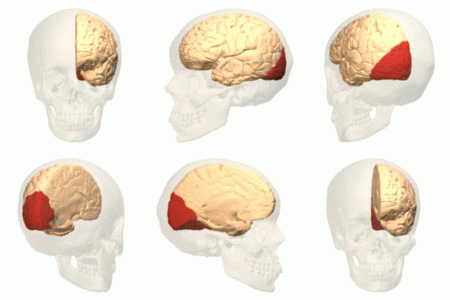
part of the brain responsible for vision | |||||
| Upload media | |||||
| Instance of |
| ||||
|---|---|---|---|---|---|
| Subclass of |
| ||||
| Part of | |||||
| Has part(s) | |||||
| |||||
Subcategories
This category has the following 16 subcategories, out of 16 total.
B
- Brodmann area 18 (16 F)
- Brodmann area 19 (14 F)
C
- Cerebral fossa (15 F)
- Cuneus (22 F)
H
L
- Lateral occipital gyrus (9 F)
- Lateral occipital sulcus (1 F)
- Lunate sulcus (2 F)
O
- Occipital pole (6 F)
P
- Pericalcarine cortex (1 F)
T
V
- Visual area V4 (18 F)
Media in category "Occipital lobe"
The following 28 files are in this category, out of 28 total.
-
Cerebrum - occipital lobe - animation.gif 320 × 320; 1.48 MB
-
Cerebrum - occipital lobe - anterior view.png 900 × 900; 284 KB
-
Cerebrum - occipital lobe - inferior view animation.gif 320 × 320; 1.77 MB
-
Cerebrum - occipital lobe - inferior view.png 900 × 900; 352 KB
-
Cerebrum - occipital lobe - lateral view.png 900 × 900; 285 KB
-
Cerebrum - occipital lobe - posterior view.png 900 × 900; 256 KB
-
Cerebrum - occipital lobe - superior view.png 900 × 900; 372 KB
-
-
-
Face post lobe occipit.png 606 × 446; 318 KB
-
Gray726 occipital lobe.png 992 × 573; 183 KB
-
Gray727 occipital lobe.png 1,025 × 598; 140 KB
-
Horizontal sections of fetal brain.jpg 960 × 720; 123 KB
-
OccCapts.png 1,662 × 781; 847 KB
-
OccCaptsLateral.png 706 × 688; 300 KB
-
OccCaptsMedial.png 879 × 766; 436 KB
-
Occipital lobe - animation.gif 320 × 320; 1.56 MB
-
Occipital lobe - inferior view animation.gif 320 × 320; 1.74 MB
-
Occipital lobe - inferior view.png 900 × 900; 421 KB
-
Occipital lobe - lateral view.png 900 × 900; 443 KB
-
Occipital lobe - posterior view.png 900 × 900; 373 KB
-
Occipital lobe - superior view.png 900 × 900; 399 KB
-
Occipital lobe animation small.gif 150 × 150; 490 KB
-
Occipital lobe in orangutan (K. Brodmann, 1909, p. 221, Fig. 136-137).jpg 1,402 × 1,098; 287 KB
-
Occipital lobe.gif 600 × 600; 4.22 MB
-
Parcellation of different cortical regions involved in visual processing.jpg 1,800 × 1,079; 205 KB
-
-
Standard anatomical parcellation of the posterior cortical surface.png 1,810 × 1,717; 1.26 MB


