Category:Centrosome
Aller à la navigation
Aller à la recherche
English: In cell biology, the centrosome is an organelle that serves as the main microtubule organizing center (MTOC) of the animal cell as well as a regulator of cell-cycle progression. It was discovered by Edouard Van Beneden in 1883
centre cellulaire organisateur des microtubules -en.svg) | |||||
| Téléverser des médias | |||||
| Nature de l’élément |
| ||||
|---|---|---|---|---|---|
| Sous-classe de |
| ||||
| Découvert(e) ou inventé(e) par |
| ||||
| Comprend |
| ||||
| |||||
Sous-catégories
Cette catégorie comprend 3 sous-catégories, dont les 3 ci-dessous.
- SVG centrosome (21 F)
C
- Centrioles (22 F)
Média dans la catégorie « Centrosome »
Cette catégorie comprend 72 fichiers, dont les 72 ci-dessous.
-
 3D-SIM-3 Prophase 3 color.jpg 902 × 896 ; 386 kio
-
-
 Anaphase.jpg 170 × 160 ; 24 kio
-
Asymmetry-of-Early-Endosome-Distribution-in-C.-elegans-Embryos-pone.0000493.s001.ogv 7,0 s, 548 × 198 ; 3,11 Mio
-
-
-
-
-
-
-
-
-
-
-
-
-
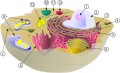 Biological cell.svg 1 466 × 891 ; 249 kio
-
Cadherin-2-Controls-Directional-Chain-Migration-of-Cerebellar-Granule-Neurons-pbio.1000240.s003.ogv 16 s, 320 × 240 ; 157 kio
-
Cadherin-2-Controls-Directional-Chain-Migration-of-Cerebellar-Granule-Neurons-pbio.1000240.s004.ogv 26 s, 320 × 240 ; 459 kio
-
Cadherin-2-Controls-Directional-Chain-Migration-of-Cerebellar-Granule-Neurons-pbio.1000240.s005.ogv 44 s, 320 × 240 ; 537 kio
-
Cadherin-2-Controls-Directional-Chain-Migration-of-Cerebellar-Granule-Neurons-pbio.1000240.s006.ogv 36 s, 320 × 240 ; 469 kio
-
Cadherin-2-Controls-Directional-Chain-Migration-of-Cerebellar-Granule-Neurons-pbio.1000240.s007.ogv 47 s, 320 × 240 ; 269 kio
-
Cadherin-2-Controls-Directional-Chain-Migration-of-Cerebellar-Granule-Neurons-pbio.1000240.s008.ogv 10 s, 1 182 × 390 ; 231 kio
-
Cadherin-2-Controls-Directional-Chain-Migration-of-Cerebellar-Granule-Neurons-pbio.1000240.s009.ogv 1 min 17 s, 320 × 240 ; 769 kio
-
Cadherin-2-Controls-Directional-Chain-Migration-of-Cerebellar-Granule-Neurons-pbio.1000240.s010.ogv 1 min 12 s, 320 × 240 ; 1,01 Mio
-
Cadherin-2-Controls-Directional-Chain-Migration-of-Cerebellar-Granule-Neurons-pbio.1000240.s011.ogv 1 min 55 s, 324 × 324 ; 338 kio
-
Cadherin-2-Controls-Directional-Chain-Migration-of-Cerebellar-Granule-Neurons-pbio.1000240.s012.ogv 11 s, 320 × 240 ; 94 kio
-
Cadherin-2-Controls-Directional-Chain-Migration-of-Cerebellar-Granule-Neurons-pbio.1000240.s013.ogv 8,8 s, 320 × 240 ; 81 kio
-
Cadherin-2-Controls-Directional-Chain-Migration-of-Cerebellar-Granule-Neurons-pbio.1000240.s014.ogv 21 s, 334 × 334 ; 92 kio
-
Cadherin-2-Controls-Directional-Chain-Migration-of-Cerebellar-Granule-Neurons-pbio.1000240.s015.ogv 18 s, 240 × 254 ; 64 kio
-
Cadherin-2-Controls-Directional-Chain-Migration-of-Cerebellar-Granule-Neurons-pbio.1000240.s016.ogv 1 min 8 s, 320 × 240 ; 166 kio
-
Cadherin-2-Controls-Directional-Chain-Migration-of-Cerebellar-Granule-Neurons-pbio.1000240.s017.ogv 11 s, 320 × 240 ; 41 kio
-
 Centrosom.jpg 265 × 184 ; 5 kio
-
 Centrosome (PSF).jpg 936 × 556 ; 94 kio
-
 Centrosome - Two centrioles -- Smart-Servier.jpg 10 240 × 5 760 ; 1,82 Mio
-
 Centrosome 1 - Two centrioles -- Smart-Servier.png 1 589 × 1 322 ; 696 kio
-
 Centrosome 2 -- Smart-Servier.png 1 177 × 1 689 ; 666 kio
-
Centrosome migration in a female Drosophila germline stem cell - journal.pbio.1001357.ogv 1 min 26 s, 560 × 420 ; 1,32 Mio
-
Centrosome migration in a living Drosophila female interphase germline stem cell - pbio.1001357.s006.ogv 4,3 s, 512 × 512 ; 38 kio
-
 CicloCentrosoma.jpg 1 020 × 733 ; 252 kio
-
 Cytokinesis-electron-micrograph.jpg 745 × 451 ; 200 kio
-
DAAM1-Is-a-Formin-Required-for-Centrosome-Re-Orientation-during-Cell-Migration-pone.0013064.s004.ogv 8,0 s, 1 036 × 684 ; 3,83 Mio
-
DAAM1-Is-a-Formin-Required-for-Centrosome-Re-Orientation-during-Cell-Migration-pone.0013064.s005.ogv 8,0 s, 1 036 × 702 ; 3,36 Mio
-
 Diplosoma.jpg 430 × 475 ; 29 kio
-
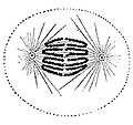
 EB1911 Cytology - centrosomes.jpg 794 × 411 ; 118 kio
-
 Gray2.png 376 × 600 ; 19 kio
-
Histone-Deacetylase-8-Is-Required-for-Centrosome-Cohesion-and-Influenza-A-Virus-Entry-ppat.1002316.s008.ogv 1,6 s, 940 × 470 ; 722 kio
-
 Interphase.png 159 × 152 ; 36 kio
-
Microtubules-are-organized-independently-of-the-centrosome-in-Drosophila-neurons-1749-8104-6-38-S1.ogv 7,4 s, 1 040 × 512 ; 4,26 Mio
-
Microtubules-are-organized-independently-of-the-centrosome-in-Drosophila-neurons-1749-8104-6-38-S5.ogv 5,7 s, 1 024 × 512 ; 856 kio
-
Microtubules-are-organized-independently-of-the-centrosome-in-Drosophila-neurons-1749-8104-6-38-S7.ogv 26 s, 512 × 453 ; 2,55 Mio
-
 Molly Sheehan Wikipedia 1.jpg 804 × 683 ; 217 kio
-
-
-
-
-
-
 Prometaphase 1.jpg 160 × 160 ; 22 kio
-
 Prophase.jpg 160 × 160 ; 20 kio
-
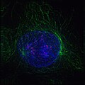 ProphaseIF.jpg 640 × 640 ; 301 kio
-
 Schanaphase.png 574 × 694 ; 401 kio
-
 Schmetaphase.png 644 × 658 ; 387 kio
-
 Schprophase.jpg 709 × 670 ; 70 kio
-
 Stages of late M phase in a vertebrate cell.svg 512 × 1 593 ; 211 kio
-
 Telophase inverse.jpg 180 × 160 ; 13 kio
-
 Telophase.jpg 180 × 160 ; 22 kio
-
The-Role-of-γ-Tubulin-in-Centrosomal-Microtubule-Organization-pone.0029795.s003.ogv 12 s, 475 × 449 ; 8,31 Mio
-
The-Role-of-γ-Tubulin-in-Centrosomal-Microtubule-Organization-pone.0029795.s004.ogv 12 s, 455 × 439 ; 8,86 Mio
-
The-Role-of-γ-Tubulin-in-Centrosomal-Microtubule-Organization-pone.0029795.s005.ogv 8,7 s, 728 × 268 ; 4,74 Mio
-
The-Role-of-γ-Tubulin-in-Centrosomal-Microtubule-Organization-pone.0029795.s006.ogv 12 s, 924 × 280 ; 4,83 Mio
-
The-Role-of-γ-Tubulin-in-Centrosomal-Microtubule-Organization-pone.0029795.s007.ogv 12 s, 439 × 448 ; 5,97 Mio
-


.jpg)


