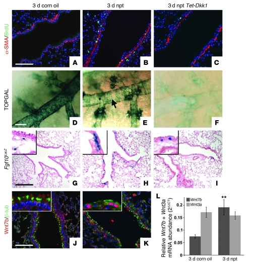
Figure 1. Wnt7b expressed by surviving ciliated cells induces Fgf10 expression in PSMCs 3 days after naphthalene-mediated Clara cell injury.
(A–C) Immunostaining for proliferation marker BrdU and SMC marker α-SMA on 2-month-old WT lungs 3 days after corn oil treatment (A), WT lungs 3 days after naphthalene (npt) treatment (B), and Rosa26-rtTa;Tet-Dkk1 lungs 3 days after naphthalene treatment (C). (D–F) β-gal staining on 2-month-old TOPGAL lungs 3 days after corn oil treatment (D), TOPGAL lungs 3 days after naphthalene treatment (E), and Rosa26-rtTa;Tet-Dkk1;TOPGAL lungs 3 days after naphthalene treatment (F). Arrow in E denotes TOPGAL activation in PSMCs. (G–I) β-gal staining on 2-month-old Fgf10LacZ lungs 3 days after corn oil treatment (G), Fgf10LacZ lungs 3 days after naphthalene treatment (H), and Rosa26-rtTa;Tet-Dkk1;Fgf10LacZ lungs 3 days after naphthalene treatment (I). Insets are enlarged ×4. Note that we identified the blue cells in alveolar compartment as lipofibroblasts (S.P. De Langhe, unpublished observations). (J and K) Immunostaining for ciliated cell marker β-tubulin and Wnt7b on 2-month-old WT lungs 3 days after corn oil treatment (J) or naphthalene treatment (K). Insets are enlarged ×3. (L) qPCR analysis of relative Wnt7b and Wnt3a mRNA abundance in 2-month-old WT lungs 3 days after treatment with corn oil versus naphthalene. **P < 0.01 vs. respective control. n = 3. Scale bars: 100 μm (A–C, J, and K); 250 μm (D–F); 200 μm (G–I).

