
Extensor retinaculum of the hand: Difference between revisions
No edit summary |
ClueBot NG (talk | contribs) m Reverting possible vandalism by 184.91.198.207 to version by Anatomist90. False positive? Report it. Thanks, ClueBot NG. (1969678) (Bot) |
||
| Line 16: | Line 16: | ||
The '''extensor retinaculum''' ('''dorsal carpal ligament''', or '''posterior annular ligament''') is an anatomical term for the thickened part of the [[antebrachial fascia]] that holds the tendons of the extensor muscles in place. It is located on the back of the [[forearm]], just [[Proximal#Directions:_general_usage|proximal]] to the hand. It is continuous with the [[palmar carpal ligament]], which is located on the anterior side of the forearm. |
The '''extensor retinaculum''' ('''dorsal carpal ligament''', or '''posterior annular ligament''') is an anatomical term for the thickened part of the [[antebrachial fascia]] that holds the tendons of the extensor muscles in place. It is located on the back of the [[forearm]], just [[Proximal#Directions:_general_usage|proximal]] to the hand. It is continuous with the [[palmar carpal ligament]], which is located on the anterior side of the forearm. |
||
It is a strong, fibrous band, extending obliquely downward and |
It is a strong, fibrous band, extending obliquely downward and medialward across the back of the [[wrist]], and consisting of part of the deep fascia of the back of the forearm, strengthened by the addition of some transverse fibers. |
||
The extensor retinaculum is attached laterally to the lateral margin of the radius. However, it is not attached to the ulna medially, as the distance between these two bones varies with supination and pronation of the forearm. Instead the medial attachment is to the most medial of the carpal bones, the triquetrum (or [[triquetral bone]]) and pisiformis (or [[pisiform bone]]). The retinaculum is also attached in its passage across the wrist, to the ridges on the dorsal surface of the radius. |
The extensor retinaculum is attached laterally to the lateral margin of the radius. However, it is not attached to the ulna medially, as the distance between these two bones varies with supination and pronation of the forearm. Instead the medial attachment is to the most medial of the carpal bones, the triquetrum (or [[triquetral bone]]) and pisiformis (or [[pisiform bone]]). The retinaculum is also attached in its passage across the wrist, to the ridges on the dorsal surface of the radius. |
||
Revision as of 01:46, 28 September 2014
| Extensor retinaculum of the hand | |
|---|---|
 | |
| Details | |
| Identifiers | |
| Latin | retinaculum musculorum extensorum manus |
| TA98 | A04.6.03.010 |
| TA2 | 2546 |
| FMA | 39987 |
| Anatomical terminology | |
The extensor retinaculum (dorsal carpal ligament, or posterior annular ligament) is an anatomical term for the thickened part of the antebrachial fascia that holds the tendons of the extensor muscles in place. It is located on the back of the forearm, just proximal to the hand. It is continuous with the palmar carpal ligament, which is located on the anterior side of the forearm.
It is a strong, fibrous band, extending obliquely downward and medialward across the back of the wrist, and consisting of part of the deep fascia of the back of the forearm, strengthened by the addition of some transverse fibers.
The extensor retinaculum is attached laterally to the lateral margin of the radius. However, it is not attached to the ulna medially, as the distance between these two bones varies with supination and pronation of the forearm. Instead the medial attachment is to the most medial of the carpal bones, the triquetrum (or triquetral bone) and pisiformis (or pisiform bone). The retinaculum is also attached in its passage across the wrist, to the ridges on the dorsal surface of the radius.
Additional images
-
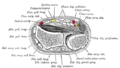 Transverse section across distal ends of radius and ulna.
 -
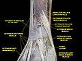 Extensor retinaculum of the hand.Deep dissection.


External links
This human musculoskeletal system article is a stub. You can help Wikipedia by expanding it. |