
Extensor retinaculum of the hand: Difference between revisions
WP:WPCHECK: Ordinal number found inside <sup> tags and other minor edits |
Among other things: removed section about attachments because it was based almost entirely on a cadaveric study (primary source). |
||
| Line 9: | Line 9: | ||
| System = |
| System = |
||
}} |
}} |
||
The '''extensor retinaculum''' ('''dorsal carpal ligament''', or '''posterior annular ligament''') is |
The '''extensor retinaculum''' ('''dorsal carpal ligament''', or '''posterior annular ligament''') is a thickened portion of the [[antebrachial fascia]] that holds the [[tendon]]s of the [[List of extensors of the human body|extensor muscles]] in place.<ref name=":2">{{Cite journal|last1=PALMER|first1=A. K.|last2=SKAHEN|first2=J. R.|last3=WERNER|first3=F. W.|last4=GLISSON|first4=R. R.|date=2016-11-17|title=The Extensor Retinaculum of The Wrist: An Anatomical and Biomechanical Study|url=https://journals.sagepub.com/doi/10.1016/S0266-7681%2885%2980006-1|journal=Journal of Hand Surgery|volume=10|issue=1|pages=11–16|language=en|doi=10.1016/S0266-7681(85)80006-1|pmid=3998587|s2cid=11507945 }}</ref> It is located on the back of the [[forearm]], just [[Proximal#Directions: general usage|proximal]] to the [[hand]].<ref name=":3">{{Cite book|last=Drake, Richard L. (Richard Lee), 1950-|url=https://www.worldcat.org/oclc/55139039|title=Gray's anatomy for students|date=2005|publisher=Elsevier/Churchill Livingstone|others=Vogl, Wayne., Mitchell, Adam W. M., Gray, Henry, 1825-1861.|isbn=0-443-06612-4|location=Philadelphia|oclc=55139039}}</ref> It is continuous with the [[palmar carpal ligament]] (which is located on the anterior side of the forearm). |
||
== Structure == |
== Structure == |
||
The extensor retinaculum is a strong, fibrous band, extending obliquely downward and medialward across the back of the [[wrist]].<ref name=":3" /> It consists of part of the [[deep fascia]] of the back of the [[forearm]], strengthened by the addition of some transverse fibers. |
The extensor retinaculum is a strong, fibrous band, extending obliquely downward and medialward across the back of the [[wrist]].<ref name=":3" /> It consists of part of the [[deep fascia]] of the back of the [[forearm]], strengthened by the addition of some transverse fibers.{{Citation needed|date=August 2023}} |
||
=== Relations === |
|||
The extensor retinaculum is attached laterally to the lateral margin of the [[radius]].<ref name=":2" /> However, it is not attached to the [[ulna]], as the distance between these two bones varies with [[Anatomical terms of motion|supination]] and [[Anatomical terms of motion|pronation]] of the [[forearm]]. Instead the medial attachment is to the [[pisiform bone]] and [[triquetral bone]].<ref name="Ryan 2011">{{cite book |last1=Ryan |first1=Stephanie |title=Anatomy for diagnostic imaging |date=2011 |publisher=Elsevier Ltd |isbn=9780702029714 |pages=256–257 |edition=Third |chapter=Chapter 7}}</ref> Other authors may state the medial attachment of extensor retinaculum with the [[fifth metacarpal bone]] and the [[pisometacarpal ligament]].<ref name=":2" /> The retinaculum is also attached in its passage across the wrist, to the ridges on the dorsal surface of the radius.<ref name=":2" /> |
|||
| ⚫ | There are six separate synovial sheaths run beneath the extensor retinaculum: (1st) abductor pollicis longus and extensor pollicis brevis tendons, (2nd) extensor carpi radialis lungus and brevis tendons, (3rd) extensor pollicis longus tendon, (4th) extensor digitorium communis and extensor indicis proprius tendons, (5th) extensor digiti minimi tendon and (6th) extensor carpi ulnaris tendon.<ref>{{Cite book |url=https://www.worldcat.org/oclc/1132300315 |title=Sobotta anatomy textbook |date=2019 |others=Friedrich Paulsen, Tobias M. Böckers, J. Waschke, Stephan Winkler, Katja Dalkowski, Jörg Mair, Sonja Klebe, Elsevier ClinicalKey |isbn=978-0-7020-6760-0 |location=Munich |oclc=1132300315}}</ref>{{Clarification needed|reason=What is the significance of the numbering? Does it signify a sequence in the mediolateral axis?|date=August 2023}} |
||
On the dorsal side of the hand, the [[palmar carpal ligament]] corresponds in location and structure to the extensor retinaculum, both being formations of the antebrachial fascia and therefore continuous. Consequently, the [[Flexor retinaculum of the hand|flexor retinaculum]] is commonly referred to as the ''transverse carpal ligament'' to avoid confusion.<ref name=":02">{{Cite book |last=Moore |first=Keith L. |title=Clinically Oriented Anatomy |last2=Dalley |first2=Arthur F. |last3=Agur |first3=Anne M. R. |date= |publisher=Wolters Kluwer |year=2018 |isbn=978-1-4963-4721-3 |edition=8th |location= |pages=159}}</ref> |
|||
| ⚫ | There are six separate synovial sheaths run beneath the extensor retinaculum: (1st) abductor pollicis longus and extensor pollicis brevis tendons, (2nd) extensor carpi radialis lungus and brevis tendons, (3rd) extensor pollicis longus tendon, (4th) extensor digitorium communis and extensor indicis proprius tendons, (5th) extensor digiti minimi tendon and (6th) extensor carpi ulnaris tendon.<ref>{{Cite book |url=https://www.worldcat.org/oclc/1132300315 |title=Sobotta anatomy textbook |date=2019 |others=Friedrich Paulsen, Tobias M. Böckers, J. Waschke, Stephan Winkler, Katja Dalkowski, Jörg Mair, Sonja Klebe, Elsevier ClinicalKey |isbn=978-0-7020-6760-0 |location=Munich |oclc=1132300315}}</ref> |
||
== Histology == |
=== Histology === |
||
Structurally, the retinaculum consists of three layers. The deepest layer, the gliding layer, consists of hyaluronic acid-secreting cells.<ref name=":0">{{Cite journal|last1=Klein|first1=David M.|last2=Katzman|first2=Barry M.|last3=Mesa|first3=Joseph A.|last4=Lipton|first4=Jeffrey F.|last5=Caligiuri|first5=Daniel A.|date=1999-01-01|title=Histology of the Extensor Retinaculum of the Wrist and the Ankle|url=http://www.sciencedirect.com/science/article/pii/S0363502399685963|journal=The Journal of Hand Surgery|language=en|volume=24|issue=4|pages=799–802|doi=10.1053/jhsu.1999.0799 |
Structurally, the retinaculum consists of three layers. The deepest layer, the gliding layer, consists of hyaluronic acid-secreting cells. The thick middle layer consists of interspersed elastin fibers, collagen bundles, and fibroblasts. The most superficial layer is made up of loose connective tissue which contains vascular channels. Combined these three layers create a smooth gliding surface as well as mechanically strong tissue which prevents tendon bowstringing.<ref name=":0">{{Cite journal |last1=Klein |first1=David M. |last2=Katzman |first2=Barry M. |last3=Mesa |first3=Joseph A. |last4=Lipton |first4=Jeffrey F. |last5=Caligiuri |first5=Daniel A. |date=1999-01-01 |title=Histology of the Extensor Retinaculum of the Wrist and the Ankle |url=http://www.sciencedirect.com/science/article/pii/S0363502399685963 |journal=The Journal of Hand Surgery |language=en |volume=24 |issue=4 |pages=799–802 |doi=10.1053/jhsu.1999.0799 |issn=0363-5023 |pmid=10447172}}</ref> The extensor retinaculum of the foot has similar structure.{{Citation needed|date=August 2023}} |
||
==Clinical significance == |
==Clinical significance == |
||
Revision as of 10:43, 6 August 2023
| Extensor retinaculum of the hand | |
|---|---|
 | |
| Details | |
| Identifiers | |
| Latin | retinaculum musculorum extensorum manus |
| TA98 | A04.6.03.010 |
| TA2 | 2546 |
| FMA | 39987 |
| Anatomical terminology | |
The extensor retinaculum (dorsal carpal ligament, or posterior annular ligament) is a thickened portion of the antebrachial fascia that holds the tendons of the extensor muscles in place.[1] It is located on the back of the forearm, just proximal to the hand.[2] It is continuous with the palmar carpal ligament (which is located on the anterior side of the forearm).
Structure
The extensor retinaculum is a strong, fibrous band, extending obliquely downward and medialward across the back of the wrist.[2] It consists of part of the deep fascia of the back of the forearm, strengthened by the addition of some transverse fibers.[citation needed]
Relations
There are six separate synovial sheaths run beneath the extensor retinaculum: (1st) abductor pollicis longus and extensor pollicis brevis tendons, (2nd) extensor carpi radialis lungus and brevis tendons, (3rd) extensor pollicis longus tendon, (4th) extensor digitorium communis and extensor indicis proprius tendons, (5th) extensor digiti minimi tendon and (6th) extensor carpi ulnaris tendon.[3][clarification needed]
On the dorsal side of the hand, the palmar carpal ligament corresponds in location and structure to the extensor retinaculum, both being formations of the antebrachial fascia and therefore continuous. Consequently, the flexor retinaculum is commonly referred to as the transverse carpal ligament to avoid confusion.[4]
Histology
Structurally, the retinaculum consists of three layers. The deepest layer, the gliding layer, consists of hyaluronic acid-secreting cells. The thick middle layer consists of interspersed elastin fibers, collagen bundles, and fibroblasts. The most superficial layer is made up of loose connective tissue which contains vascular channels. Combined these three layers create a smooth gliding surface as well as mechanically strong tissue which prevents tendon bowstringing.[5] The extensor retinaculum of the foot has similar structure.[citation needed]
Clinical significance
Studies conducted on the retinaculum have exhibited it to have several possible surgical treatments uses. A graft of the extensor retinaculum was shown to be useful in treating boxer's knuckle when direct repair of the damaged capsule is not possible.[6] Because of their similarities in histological structure, studies also show the extensor retinaculum to be a reasonable biological replacement for reconstruction of a deficient annular pulley.[6]
Additional images
-
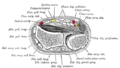 Transverse section across distal ends of radius and ulna.
 -
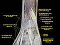 Extensor retinaculum of the hand.Deep dissection.


References
- ^ PALMER, A. K.; SKAHEN, J. R.; WERNER, F. W.; GLISSON, R. R. (2016-11-17). "The Extensor Retinaculum of The Wrist: An Anatomical and Biomechanical Study". Journal of Hand Surgery. 10 (1): 11–16. doi:10.1016/S0266-7681(85)80006-1. PMID 3998587. S2CID 11507945.
- ^ a b Drake, Richard L. (Richard Lee), 1950- (2005). Gray's anatomy for students. Vogl, Wayne., Mitchell, Adam W. M., Gray, Henry, 1825-1861. Philadelphia: Elsevier/Churchill Livingstone. ISBN 0-443-06612-4. OCLC 55139039.
{{cite book}}: CS1 maint: multiple names: authors list (link) CS1 maint: numeric names: authors list (link) - ^ Sobotta anatomy textbook. Friedrich Paulsen, Tobias M. Böckers, J. Waschke, Stephan Winkler, Katja Dalkowski, Jörg Mair, Sonja Klebe, Elsevier ClinicalKey. Munich. 2019. ISBN 978-0-7020-6760-0. OCLC 1132300315.
{{cite book}}: CS1 maint: location missing publisher (link) CS1 maint: others (link) - ^ Moore, Keith L.; Dalley, Arthur F.; Agur, Anne M. R. (2018). Clinically Oriented Anatomy (8th ed.). Wolters Kluwer. p. 159. ISBN 978-1-4963-4721-3.
- ^ Klein, David M.; Katzman, Barry M.; Mesa, Joseph A.; Lipton, Jeffrey F.; Caligiuri, Daniel A. (1999-01-01). "Histology of the Extensor Retinaculum of the Wrist and the Ankle". The Journal of Hand Surgery. 24 (4): 799–802. doi:10.1053/jhsu.1999.0799. ISSN 0363-5023. PMID 10447172.
- ^ a b Nagaoka, Masahiro; Satoh, Takako; Nagao, Soya; Matsuzaki, Hiromi (2006-07-01). "Extensor Retinaculum Graft for Chronic Boxer's Knuckle". The Journal of Hand Surgery. 31 (6): 947–951. doi:10.1016/j.jhsa.2006.02.027. ISSN 0363-5023. PMID 16843154.